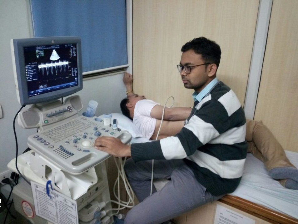
2D Echo
2D echo, is a non-invasive test used to analyze the functioning and assess the sections of your heart. This test gives images of the different parts of the heart with the help of sound vibrations.It assists in checking damages, blockages, and blood flow rate.
Why should you undergo a 2D echo?
2D Echo is done to detect the following heart conditions:
- Any underlying heart diseases or abnormalities
- Congenital heart diseases and blood clots or tumors
- Malfunctioning of the heart valve
- Abnormality of blood flow within the heart

CT Scan
A computed tomography scan (CT scan) is a medical imaging technique used to obtain detailed internal images of the body.
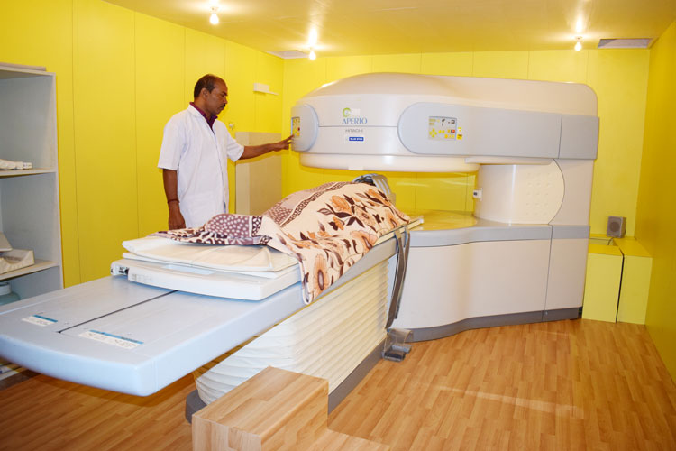
MRI
Magnetic resonance imaging (MRI) is a medical imaging technique used in radiology to form pictures of the anatomy and the physiological processes inside the body. MRI scanners use strong magnetic fields, magnetic field gradients, and radio waves to generate images of the organs in the body. MRI does not involve X-rays or the use of ionizing radiation, which distinguishes it from computed tomography (CT) and positron emission tomography (PET) scans.MRI is widely used in hospitals and clinics for medical diagnosis, staging and follow-up of disease. Compared to CT, MRI provides better contrast in images of soft tissues, e.g. in the brain or abdomen.

EEG
An electroencephalogram (EEG) is a test that measures electrical activity in the brain using small, metal discs (electrodes) attached to the scalp. Brain cells communicate via electrical impulses and are active all the time, even during asleep. This activity shows up as wavy lines on an EEG recording.An EEG is one of the main diagnostic tests for epilepsy. An EEG can also play a role in diagnosing other brain disorders.

Treadmill Test
A treadmill test, also known as a TMT test or exercise stress test, involves walking or running on a treadmill. This test is used to assess how effectively the heart responds when it is working harder than it does at rest. The TMT test serves as a medical diagnostic tool, aiding in the evaluation of heart function for individuals with various health conditions that may predispose them to a higher risk of heart diseases. A TMT test is an evaluation procedure to assess overall heart health. During this test, patients are evaluated based on how far they can run on a treadmill before experiencing irregular heart rhythms or reduced blood supply to the heart.
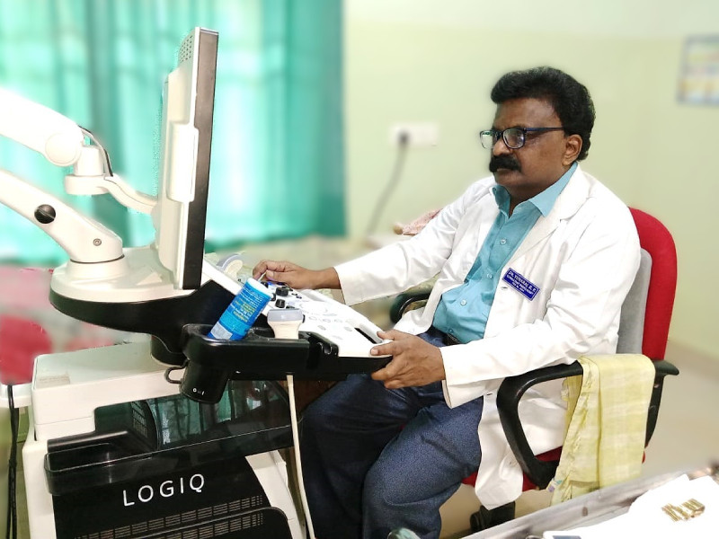
Ultrasonography
Dr. Yuvaraj R P is a well qualified and skilled radiologist. The ultrasound department functions under his experienced hands. He is available from Monday to Saturday at 9:00am to 3:00pm. We have an advanced Ultrasound machine Wipro GE( Logiq P9R3). It is used to create an image of internal body structures such as tendons, muscles, joints, blood vessels, and internal organs, to measure some characteristics (e.g. distances and velocities) and in examining pregnant women.

X-ray
X-ray diagnostic imaging is a method for taking pictures of areas inside the body. It uses a small amount of radiation to produce pictures of the body’s internal structures. X-rays are the oldest and most frequently used form of medical imaging. They are often used to help diagnose fractured bones, look for injury or infection, and to locate foreign objects in soft tissue. The X-rays pass through the body, creating an image on film or a computer display.
Laboratory services
The Laboratory at St. Ignatius Hospital is a vital component of our healthcare system, offering comprehensive diagnostic services to support patient care. Equipped with advanced technology and staffed by skilled professionals, the laboratory provides a wide range of services, including Hematology, Biochemistry, Microbiology and Serology, Pathology, Histopathology and Cytopathology. These services enable accurate diagnosis and effective monitoring of various medical conditions. The Hematology section conducts tests to assess blood disorders, while the Biochemistry department analyzes bodily fluids to evaluate organ function and metabolic health. Microbiology and Serology focus on identifying infectious agents, and Pathology, Histopathology, and Cytopathology provide essential insights into tissue and cellular changes, crucial for diagnosing diseases. With a commitment to precision and timely results, the Laboratory at St. Ignatius Hospital plays a key role in enhancing patient outcomes and facilitating informed treatment decisions.
ECT
Electroconvulsive therapy (ECT) or electroshock therapy (EST) is a psychiatric treatment where a generalized seizure (without muscular convulsions) is electrically induced to manage refractory mental disorders. ECT is often used as an intervention for major depressive disorder, mania, and catatonia
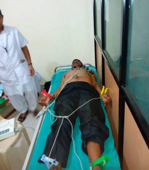
ECG
Electrocardiography is the process of producing an electrocardiogram, a recording of the heart’s electrical activity through repeated cardiac cycles. These electrodes detect the small electrical changes that are a consequence of cardiac muscle depolarization followed by repolarization during each cardiac cycle (heartbeat). Changes in the normal ECG pattern occur in numerous cardiac abnormalities, including:Cardiac rhythm disturbances (such as atrial fibrillation and ventricular tachycardia),Inadequate coronary artery blood flow (such as myocardial ischemia and myocardial infarction) and electrolyte disturbances, such as hypokalemia.
2D Echo
2D echo, is a non-invasive test used to analyze the functioning and assess the sections of your heart. This test gives images of the different parts of the heart with the help of sound vibrations.It assists in checking damages, blockages, and blood flow rate.
Why should you undergo a 2D echo?
2D Echo is done to detect the following heart conditions:
- Any underlying heart diseases or abnormalities
- Congenital heart diseases and blood clots or tumors
- Malfunctioning of the heart valve
- Abnormality of blood flow within the heart
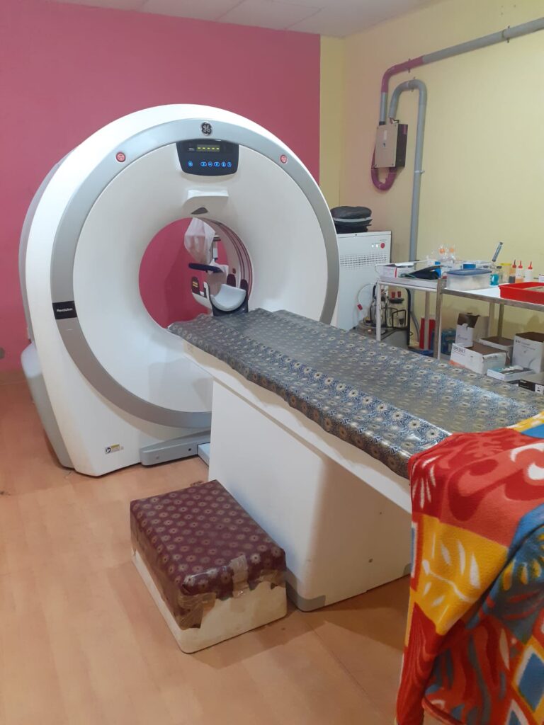
CT Scan
A computed tomography scan (CT scan) is a medical imaging technique used to obtain detailed internal images of the body.
MRI
Magnetic resonance imaging (MRI) is a medical imaging technique used in radiology to form pictures of the anatomy and the physiological processes inside the body. MRI scanners use strong magnetic fields, magnetic field gradients, and radio waves to generate images of the organs in the body. MRI does not involve X-rays or the use of ionizing radiation, which distinguishes it from computed tomography (CT) and positron emission tomography (PET) scans.MRI is widely used in hospitals and clinics for medical diagnosis, staging and follow-up of disease. Compared to CT, MRI provides better contrast in images of soft tissues, e.g. in the brain or abdomen.
EEG
An electroencephalogram (EEG) is a test that measures electrical activity in the brain using small, metal discs (electrodes) attached to the scalp. Brain cells communicate via electrical impulses and are active all the time, even during asleep. This activity shows up as wavy lines on an EEG recording.An EEG is one of the main diagnostic tests for epilepsy. An EEG can also play a role in diagnosing other brain disorders.
Treadmill Test
A treadmill test, also known as a TMT test or exercise stress test, involves walking or running on a treadmill. This test is used to assess how effectively the heart responds when it is working harder than it does at rest. The TMT test serves as a medical diagnostic tool, aiding in the evaluation of heart function for individuals with various health conditions that may predispose them to a higher risk of heart diseases. A TMT test is an evaluation procedure to assess overall heart health. During this test, patients are evaluated based on how far they can run on a treadmill before experiencing irregular heart rhythms or reduced blood supply to the heart.
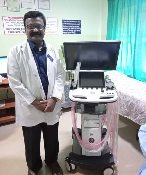
Ultrasonography
Dr. Yuvaraj R P is a well qualified and skilled radiologist. The ultrasound department functions under his experienced hands. He is available from Monday to Saturday at 9:00am to 3:00pm. We have an advanced Ultrasound machine Wipro GE( Logiq P9R3). It is used to create an image of internal body structures such as tendons, muscles, joints, blood vessels, and internal organs, to measure some characteristics (e.g. distances and velocities) and in examining pregnant women.
X-ray
X-ray diagnostic imaging is a method for taking pictures of areas inside the body. It uses a small amount of radiation to produce pictures of the body’s internal structures. X-rays are the oldest and most frequently used form of medical imaging. They are often used to help diagnose fractured bones, look for injury or infection, and to locate foreign objects in soft tissue. The X-rays pass through the body, creating an image on film or a computer display.
ECT
Electroconvulsive therapy (ECT) or electroshock therapy (EST) is a psychiatric treatment where a generalized seizure (without muscular convulsions) is electrically induced to manage refractory mental disorders. ECT is often used as an intervention for major depressive disorder, mania, and catatonia
ECG
Electrocardiography is the process of producing an electrocardiogram, a recording of the heart’s electrical activity through repeated cardiac cycles. These electrodes detect the small electrical changes that are a consequence of cardiac muscle depolarization followed by repolarization during each cardiac cycle (heartbeat). Changes in the normal ECG pattern occur in numerous cardiac abnormalities, including:Cardiac rhythm disturbances (such as atrial fibrillation and ventricular tachycardia),Inadequate coronary artery blood flow (such as myocardial ischemia and myocardial infarction) and electrolyte disturbances, such as hypokalemia.